1.1.4 Ultrasound (US): Sound Waves & Echoes
“Real-time imaging turned diagnosis into something you can watch.” — Why ultrasound is everywhere in clinical practice
After CT, MRI, and X-ray (the “anatomical imaging trio”), ultrasound stands out as the real-time, radiation-free, portable, and cost-effective modality that clinicians reach for first in many scenarios.
🔊 From SONAR to medical imaging
Early roots
Ultrasound (frequency > 20 kHz) was first widely used in SONAR for submarine detection in World War I. Later, it became a workhorse in industrial nondestructive testing.
Bringing it into medicine was harder: biological tissues are complex, and clinicians need fast and repeatable imaging.
Clinical pioneers
- 1942: Karl Dussik attempted ultrasound transmission imaging for brain tumors (limited by the skull).
- 1958: Ian Donald demonstrated obstetric ultrasound and helped establish medical ultrasound as a clinical tool.
📡 Imaging principle: echoes + piezoelectric transducers
Frequency vs penetration
Medical ultrasound typically operates at 1–20 MHz:
- Low frequency (1–5 MHz): deeper penetration (abdomen)
- High frequency (7–20 MHz): higher resolution (thyroid, vessels)
Piezoelectric effect
The transducer uses piezoelectric crystals (e.g., PZT):
- Apply voltage → crystal vibrates → emits ultrasound
- Echo returns → crystal vibrates → generates voltage (signal)
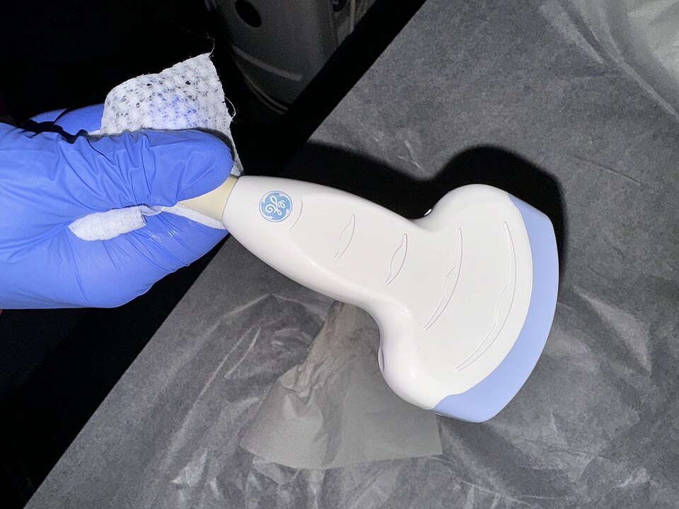 A linear-array ultrasound probe with multiple piezoelectric elements
Interaction with tissue
- Reflection: the main information source; depends on acoustic impedance mismatch
- Scattering: creates speckle/texture
- Attenuation: increases with depth and frequency
- Refraction: may cause artifacts
Common modes
| Mode | What it shows | Typical use |
|---|---|---|
| B-mode | 2D grayscale | general imaging |
| M-mode | motion over time | cardiac valves |
| Color Doppler | flow overlay | vascular / cardiac |
Doppler equation
Blood flow velocity can be estimated by:
💡 Why gel is required
Air has a very different acoustic impedance than tissue, causing near-total reflection at the air–skin interface. The coupling gel removes air gaps so ultrasound energy can enter the body.
🚀 Technology evolution (high-level)
| Era | Key breakthroughs | Typical impact |
|---|---|---|
| 1970s–1980s | Real-time B-mode | true dynamic imaging |
| 1980s–1990s | Doppler | flow/hemodynamics |
| 1990s–2000s | 3D/4D, harmonic imaging | better visualization & contrast |
| 2000s–2010s | Contrast US, elastography | functional assessment |
| 2010s–today | AI + handheld devices | accessibility & workflow |
Where to go next
- PET/SPECT:
1.1.5 PET/SPECT: Metabolism & Function(/en/guide/ch01/01-modalities/05-pet) - Practical preprocessing topics (attenuation correction / denoising): Chapter 2.3 (
/en/guide/ch02/03-pet-us-preprocessing)